
This post was contributed by Andrew Youngson, media and publicity officer in Birkbeck External Relations.
During Arts Week, former Birmingham poet laureate Stephen Morrison-Burke, was announced as the inaugural recipient of the Kit de Waal scholarship – a creative writing scholarship specially designed for budding writers who would not otherwise be able to afford a Master’s degree.
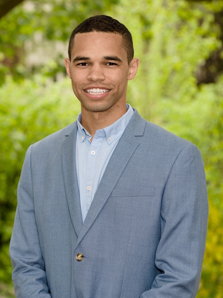
Stephen Morrison-Burke
A few years ago, Stephen’s motivation to write poetry began to give way to a new writing urge: to write prose. The result is his debut novel, The Purple Sun – a semi-biographical tale inspired by his father’s experiences leaving his Jamaican homeland in the 1970s to begin a new life in the UK. This month, Stephen finished the final draft, an 90,000-word manuscript, which follows two-and-a-half years of writing, primarily in the very early mornings. (For the full story, read the news article here)
At the Arts Week event – the Creative Writing Alumni showcase – Stephen offered his thanks for the opportunity to undertake the MA Creative Writing (part-time) programme over the next two years, then delivered a rousing rendition of a poem of his, called Wishlist.
Here, Stephen talks about the scholarship opportunity, and his relationship with writing.
Hi Stephen. Why did you decide to apply for the Kit de Waal scholarship?
“When you are essentially teaching yourself, there’s a lot you don’t learn about the theoretical elements, such as structure, plot, pace and character development, so I thought the opportunity to go through that with professionals in their fields was something I didn’t want to pass up.”
How did you feel when you were interviewed to interview for the scholarship?
“Instantly I was overjoyed. It was a very tough time for me, and it can be pretty lonely writing by yourself. So when I got that through I can’t remember feeling as relieved as that in a long time. It wasn’t necessarily that I thought I could win, it was just more that I saw an opportunity to showcase what I had been working on for so long.
Why did you decide to write a novel?
“I had no intention of writing a novel, that’s the honest truth. It sounds mad, but I just had these gut feelings that wouldn’t go. And when I started to write, I just felt better, like I was finally doing what I was supposed to be doing. I felt relieved. But it’s strange that at the exact same time as I got these feelings, the poetry stopped.
“I had had my busiest month ever in poetry – I had met the Queen, I had travelled round the country, I’d written and performed a poem for Prince William – but come New Years Eve 2013, everything just stopped, and this novel took priority. Since then, I’ve done bits and pieces with poetry, but really I’ve just focused on this novel.”
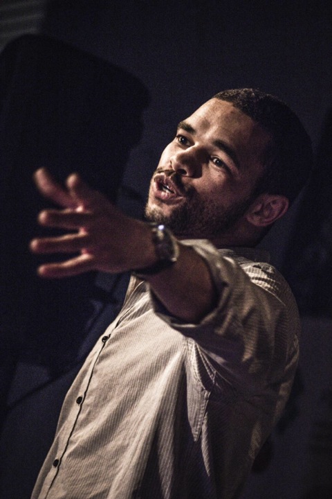
Stephen Morrison-Burke performing poetry
Poetry vs prose
“Although they are similar, I have to treat them very different. I have to respect the art form of writing novels. Strangely enough, my poetry is mostly storytelling anyway.”
Why do you choose to write at 4am?
“I’m a nightmare. If the sun’s out, I always end up procrastinating looking at my phone or on the internet. If it’s dark, there’s nothing else I can do, so there’s no other choice but to write.”
Do you get writer’s block?
“I don’t believe in writer’s block. I always believe I can write something, even if it’s nonsense, or just a short poem or something to plug the gap. But the writing is a slog, it’s hard work. There are no two ways about it. I thought it would be easier than it’s been, but I chip away at it one day at a time, one sentence at a time, one word at a time. I just turn up and make sure I’m writing something.”
Why does writing make you feel better?
“I felt like there was a lot I had to say that I wasn’t saying. There was a lot to get off my chest. I’m quite quiet and introverted, so by not getting it out it felt like it was building up. So when I was writing it was cathartic.
“From the things I had learned and experienced living in a tough part of Birmingham, to then boxing for 10 years of my life, to then all this poetry, there was a lot I wanted to say. I just wasn’t saying anything about that, so it was a relief to write it down. I thought I would only write one book and it would all come out in one go, but now that I’ve written one, I feel I could write another ten.”
How has your style developed over time?
“It’s certainly developed. It’s been a mirror of who I am as a person. I started off a little pretentious maybe, trying to impress. And certainly the poetic influence can make you embellish the writing. But the more I went along, and the more I read the likes of Hemingway, Steinbeck and Amy Hempel, the more I realised it can be straight to the point and not too airy fairy. It’s about trying to see things different to how everybody else does, which is why I’m so fascinated with the perspectives of children.”
What can you say about the background to your novel?
“It’s loosely based on a true story – my dad’s. My dad and I have been working on this together since Day One. He’s the one that said ‘you can do something with this, it’s going to be special’. He would always gee me up and gave me the motivation to see it through. It was just me on my computer, and he gave me the motivation to do something.”
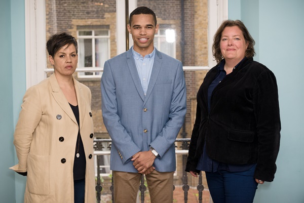
(l-r) Kit de Waal, Stephen Morrison-Burke, MA Creative Writing director Julia Bell
The latter half of the book deals with violence. What can you say about that?
“That topic is not something my Dad would go into. That’s where I had to go into my own feelings. This is where I related back to Dostoyevsky’s Crime and Punishment, and started to be creative. It’s not just violence for violence’s sake. I wanted to understand the mind behind violence, and what would drive someone who’s intelligent to turn to that life.”
Does your poetry background influence your prose writing?
“I feel I’m able to draw on it. I focus on the form of novels and sometimes the poetry will come through. For instance sometimes words come out in rhyme. I have to stop myself, but then at times I find it creates a good rhythm to the sentence when two words rhyme. So I would be very careful and selective about how I use poetry. But there is a very thin line between the two, if a line at all. So I let them wrestle between themselves.”
How does it feel when you are in the writing flow?
“Being in the flow is very rare for me, to be honest. I’d compare writing to how I imagine riding rodeo would feel like. You have to hold on as tight as you can until it throws you off, and that’s the end of your day, when you run out of juice. It could be three hours, or one or seven. You just hold on as tight as you can and afterwards you wait for the next day to come round.”
How did you find the interview for the scholarship with Julia Bell and Kit de Waal
“They gave me a lot of encouragement, the fact that I had got that far. On the day I said to them it was great to hear that I was on the right track with my writing. They said it was brilliant, which was actually the first feedback I had had on the writing. I was so happy to hear that.”
What do you want to get out of the MA Creative Writing programme?
“If I’m honest, I came into this wanting to make some kind of living through writing books. But I don’t put any pressure on the course to deliver that for me. My goal is to make a living out of writing and I know the course will help me, to say the least.”
“I really want to contextualise books. When I read them, there’s no context beyond reading the introduction, so for the lecturers to paint a picture of the times the books were written, and to talk about what was going on at social and political levels, will be really useful. As it is right now, I read a book from first chapter to the last, but with little understanding outside of the words I’ve read. So it will be great to sit down with a professional to discuss the whys and hows.”
Find out more


 Ideally, we would want to watch this, or any other form of molecular motion, in real time, but this is impossible because molecules are far too small: smaller than the wavelength of light, so they cannot be viewed in a light microscope. Studies of molecular structure require techniques like X-ray crystallography and electron microscopy, both of which have been used to study motor molecules.

 These two proteins have now also been studied using two other structural biology techniques, nuclear magnetic resonance and, most recently, electron microscopy. This last technique is best suited for studying large proteins and complexes of many protein chains, and therefore not suitable for studying most forms of haemoglobin, a small, simple protein. Haemoglobin in earthworms, however, functions as a complex of many individual molecules. Electron microscopy gave a low-resolution picture of the overall shape of these molecules, much like those first haemoglobin structures, and a more precise picture was built up by ‘docking’ atomic-resolution X-ray structures of a single haemoglobin molecule into the shape of the fold.